Mitochondrial Localization Of Viral Proteins Essay

Mitochondrial Localization Of Viral Proteins Essay

Question:

Discuss About The Mitochondrial Localization Of Viral Proteins.

Answer:

Introduction

The following is a summary of a peer review journal article. Piller et al.,(1999) compile a study titled, “The Amino-Terminal Region for Vpr from Human Immunodeficiency Virus Type 1 Forms Ion Channels and Kills Neurons.” The study is a qualitative kind of research that uses mixed methods research to gather findings. Piller et al.,(1999) seek to examine the terminal for the amino regions for management originating from human HIV.  Mitochondrial Localization Of Viral Proteins Essay.he study employs the use of mutagenesis directed to the site of infection together with synthetic peptides. Piller et al.,(1999) establishe various substantial findings. The study identifies the structural regions which are charged with the above metabolic functions.

The study posits that there have been previous attempts to report on the accessory Vpr protein from the HIV. Piller et al.,(1999) find that the channels of activity are altered by the changes due to mutations in the region of the N-terminal of the Vpr (Piller et al., 1999). The variations that occur in the hydrophobic region of the Vpr or the amino acids that range between 53 and 71 do not affect the activity of the N-terminal. Piller et al.,(1999) use the initials to mean Human resource management Immunodeficiency Virus responsible for AIDS infection (Castanier & Arnoult, 2011). The HIV type 1 virus forms channels of action-selection in planar bilayers of lipids. The virus can depolarize neurons that are intact and depolarized. It causes an inward sodium current that results in the death of cells (Castanier & Arnoult, 2011).

Piller et al.,(1999) analyzed the mutations that contain changes in the primary C terminus. The results confirmed previous findings that suggested the region was responsible for the rectification that was observed in the wild versions of the Vpr currents. Piller et al.,(1999) find that a peptide that comprises the first 40 N-terminal amino acids of Vpr is enough for the formation of a channel of ions that are the same as ions that result from the wild type of Vpr found in the planar layers of lipids. Mitochondrial Localization Of Viral Proteins Essay.

ORDER A PLAGIARISM-FREE PAPER NOW

References

Castanier, C. & Arnoult, D., 2011. Mitochondrial localization of viral proteins as a means to subvert

Viruses have developed a battery of distinct strategies to overcome the very sophisticated defense mechanisms of the infected host. Throughout the process of pathogen–host co-evolution, viruses have therefore acquired the capability to prevent host cell apoptosis because elimination of infected cells via apoptosis is one of the most ancestral defense mechanism against infection. Conversely, induction of apoptosis may favor viral dissemination as a result of the dismantlement of the infected cells. Mitochondria have been long recognized for their key role in the modulation of apoptosis but more recently, mitochondria have been shown to serve as a crucial platform for innate immune signaling as illustrated by the identification of MAVS. Thus, it is therefore not surprising that this organelle represents a recurrent target for viruses, aiming to manipulate the fate of the infected host cell or to inhibit innate immune response. In this review, we highlight the viral proteins that are specifically targeted to the mitochondria to subvert host defense. This article is part of a Special Issue entitled Mitochondria: the deadly organelle.
Mitochondria have fascinated researchers across all disciplines of life science for over a century [1]. These eukaryotic organelles most likely originate from an aerobic bacterium, related to modern α-proteobacteria, that was incorporated by the progenitor of the eukaryotic cell. Mitochondrial Localization Of Viral Proteins Essay. In the development of the endosymbiotic relationship, the bacterium retained its ring-shaped chromosome but lost most of its genes. In all eukaryotes, approximately 99% of the mitochondrial proteins are encoded by the nuclear DNA and synthesized by cytosolic ribosomes [2,3,4]. The efficient selection and uptake of newly synthesized proteins is mediated by an elaborate machinery in the mitochondrial outer and inner membranes in cooperation with several soluble factors. The decisive entry gate is formed by a complex of outer membrane proteins named TOM complex (translocase of the mitochondrial outer membrane [5]). A prominent component of the TOM complex, albeit only loosely associated, is Tom70.
The mitochondrial proteome comprises ~1000 (yeast)-1500 (human) different proteins. In the yeast Saccharomyces cerevisiae, 74 integral and 21 peripheral outer mitochondrial membrane proteins were identified by a sophisticated proteomic approach [6,7,8]. Tom70 is an abundant protein of the mitochondrial outer membrane, and in S. cerevisiae, the corresponding gene (gene: YNL121C; UniProtKB-P07213; synonym: MAS70) was identified already in 1983 [9,10,11]. The characterization of Tom70 as an import receptor for a subset of mitochondrial proteins followed several years later: Studies on the import of proteins into the mitochondria of the filamentous fungus Neurospora crassa revealed a role of Tom70 (gene: NCU04245; UniProtKB–P23231; synonym: MOM72) in binding of newly synthesized metabolite carrier proteins such as the ADP/ATP carrier of the mitochondrial inner membrane [12], and this function was confirmed for the Tom70 of yeast [13,14]. In parallel, the outer membrane protein Tom20 was identified as a receptor for precursor proteins targeted by an amino terminal presequence and Tom40 was characterized as the central pore-forming component of the TOM complex. Since then, the system of the two import receptors Tom70 and Tom20 in cooperation with the channel-forming Tom40 was regarded as the central structure the TOM complex. Mitochondrial Localization Of Viral Proteins Essay. Subsequent studies showed that the TOM complex contains several additional components and cooperates with independent complexes of other outer membrane proteins, but they essentially confirmed the basic scheme of distinct receptor proteins, one of them being Tom70, cooperating with a general import pore [3,4] (see below, Figure 5). Although Tom70 functionally cooperates with the core components of the TOM complex to facilitate import of mitochondrial proteins, it is not permanently associated with Tom40 [13,15,16,17,18,19,20]. The molecular structure of the TOM complex was recently resolved by high resolution Cryo-EM [21,22,23]. The studies confirmed the tight and stable association of Tom40 with several additional Tom proteins, but Tom70 was not included. Tom70 is thus an outer membrane protein that associates with Tom40 only partially and in a reversible manner.
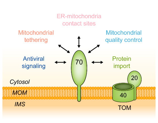
But why is it useful to review the current state of research on a protein that was characterized 30 years ago, if the basic scheme of its function seems to be retained? In fact, in recent years, a wealth of new data has emerged, showing that Tom70 has additional functions, some of them being entirely independent of the biogenesis of mitochondrial proteins: (1.) In 2006, the first high resolution crystal structure of a Tom70 was published [24]. (2.) Already in 2003, data had revealed a defined binding site within Tom70 for cytosolic Hsp70 [25]. (3.) A series of new studies on tetratricopeptide (TPR) domain-containing proteins of the endoplasmic reticulum and of chloroplasts revealed striking similarities to Tom70 both in the tertiary structure and also in the function as a receptor for heat shock proteins. (4.) In 2011, the activity of Tom70 in binding of preproteins was shown to be regulated by reversible phosphorylation of a distinct serine residue [26]. (5.) The advent of whole genome sequencing allowed a first insight into the evolution of Tom70 in different eukaryotic lineages. (6.) Tom70 was found to participate in direct interactions between mitochondria and partner proteins of the endoplasmic reticulum. (7.) TOM70 interacts with the mitochondrial antiviral-signaling protein (MAVS), a component in a system of antiviral immunity, and some data indicate that human TOM70 is a target of a protein encoded by the genome of the SARS-CoV-2 (the severe acute respiratory syndrome coronavirus 2) [27,28]. (8.) Eventually, new investigations confirm that Tom70 facilitates the mitochondrial import of many different proteins, but they also indicate that the specific selection of proteins by the TOM complex is independent of Tom70 [29]. Together, the new observations demonstrate that Tom70 is a versatile mediator in protein traffic, membrane contact sites and signaling (Figure 1). Mitochondrial Localization Of Viral Proteins Essay.
Figure 1. Cellular functions of the mitochondrial outer membrane protein Tom70. IMS, intermembrane space; MOM, mitochondrial outer membrane; TOM, translocase of the mitochondrial outer membrane, containing the channel-forming protein Tom40 and the import receptor Tom20.
2. Molecular Structure of Tom70

2.1. The Structure of Monomeric Tom70

Yeast Tom70 is a 70.1 kDa protein of 617 amino acids anchored in the mitochondrial outer membrane. The membrane anchor is located in the extreme N-terminus, which harbors a hydrophobic segment (between amino acids 9 and 38) whereas the hydrophilic receptor domain is exposed in the cytosol [11,13]. The insertion of newly synthesized Tom70 into the outer membrane is mediated by the MIM complex and proceeds independently of both Tom20 and Tom70 of the TOM complex [30,31].
A crystal structure of yeast Tom70 at a resolution of 3.0 Å was provided in a landmark publication by Wu & Sha (2006). The crystal structure revealed that Tom70 is essentially a bundle of 26 α-helices (A1–A26), of which the majority is involved in the formation of 11 tetratricopeptide repeat (TPR) motifs (Figure 2A). A Tom70 monomer forms a supra-helical structure with two distinct parts: An N-terminal domain (helices A1–A7) and a C-terminal domain (helices A8–A26) (Figure 2B). The Tom70 superhelix has a length of approximately 100 Å and a radius of 50 Å. The N-terminal and the C-terminal domain are connected to each other in a “head to head” orientation, where the C-terminal ends of both domains face each other with antiparallel helices A7 and A25 [24].
Figure 2. Overview of the structural features of S. cerevisiae Tom70. (A): Crystal structure of dimeric Tom70. Structural data were obtained from Wu & Sha (2006). Each Tom70 monomer contains 617 amino acids and has a molecular mass of approximately 70 kDa. The secondary structure of Tom70 is characterized by 26 α-helices (A1–A26) that form 11 tetratricopeptide repeat (TPR) motifs and create its supra-helical tertiary structure. The N-terminal domain (A1–A7) of Tom70 contains a chaperone binding site formed by a clamp-type TPR domain. The binding site is defined by an arginine at position 171 and a cysteine at position 141 [25,29]. The TPR clamp contains a serine at position 174 which can be phosphorylated by protein kinase A (PKA) in response to metabolic changes [26]. Within its C-terminus, Tom70 contains a highly conserved groove which displays mainly hydrophobic and a few polar residues at its top and three conserved glutamates at its bottom. Mitochondrial Localization Of Viral Proteins Essay. The opposite site of the TPR clamp domain contains a methionine at position 551 which might be important for presequence recognition by Tom70 [40]. (B): Configurations of the N-terminal domains (A1–A7) of S. cerevisiae Tom70 [24] and Tom71 [37]. The different orientations of the N-terminal domain relative to the C-terminal domain are suggested to indicate that both proteins may adopt different conformational states.
A defined chaperone binding site is contained in the N-terminal domain of Tom70: The helices A1–A6 (TPR motifs 1–3) form a clamp-type TPR domain that serves as a chaperone acceptor for Hsp70 in yeast and for Hsp70 and Hsp90 in mammals [24,25,32,33].
In parallel to Tom70, the genome of S. cerevisiae encodes a functional paralog, Tom71, which is expressed at low levels [34,35,36]. In comparison to Tom70, Tom71 was found to display a strikingly divergent arrangement of its C- and N-terminal domains. The differences seem to refer to alternative conformational states of both Tom70 and Tom71, including an open and a closed conformation (Figure 2B), and this flexibility was suggested to determine the accessibility of a binding site for protein recognition [37,38,39,40].

2.2. Putative Preprotein Binding Sites

It is traditionally assumed that Tom70 not only binds, but also selects a subset of mitochondrial preproteins by direct interactions with structures that are specific for these proteins. The essential receptor sites within the structure of Tom70 that serve this purpose are still unclear, although possible binding sites were characterized in several studies:
An early study reported a stably folded 25 kDa core domain of Tom70 (amino acids 247–460, hence outside the chaperone binding site), that was able to bind to chemically synthesized internal segments of substrate proteins in vitro with a specificity comparable to the full length receptor [41]. Mitochondrial Localization Of Viral Proteins Essay. The assays of this comprehensive study revealed a distinct pattern of affinities, but the relevance of these affinities for chaperone-mediated binding of preproteins to Tom70 in vivo was not investigated.
Analyzing the crystal structure of yeast Tom70, Wu & Sha (2006) identified a highly conserved groove located in the center of its C-terminal domain (containing TRP motifs 4–11) [24]. While the distal side of the binding groove is mainly made up of conserved hydrophobic and polar residues (hydrophobic: Pro252, Phe260, Phe341, Leu342, Met406, Phe408, Ile409, Phe432, Phe470, Ile474; polar: Asp375, Gln436, Gln405), the proximal side contains three conserved residues that are negatively charged (Glu473, Glu542, Glu577). The authors suggested this groove to represent the major binding site for preproteins, with the conserved hydrophobic parts acting as docking site for the hydrophobic substrates of Tom70 [24]. However, this function has not yet been confirmed by experimental evidence.
In a study on the role of Tom70 in the mitochondrial import of carrier proteins, Tom70 variants were included with the highly conserved glutamates in the proposed binding groove exchanged against alanine. The experiments did not show any indication of an involvement of this structure in carrier protein binding or selection [29]. An independent study found that presequence peptides can be photo-crosslinked to residues at least in close proximity to the postulated binding groove [40]. A variant of Tom70 in which the Met551 was replaced by an arginine showed reduced affinity to Mdl1 (a presequence-containing precursor) but did not impair the import of carrier proteins. The results of this study are in agreement with a possible role of Tom70 in direct interactions with presequences of precursor proteins, but the role of the postulated binding groove in these interactions is still unclear. Mitochondrial Localization Of Viral Proteins Essay.
In retrospect, the data on binding sites that may determine a specificity of Tom70 in the selection of subsets of mitochondrial preproteins do not provide a clear picture. The only site of Tom70 that unambiguously serves as a docking site for preproteins is thus the site within the N-terminal part of Tom70 that is able to bind Hsp70 and Hsp70-bound preproteins [25,29]. The function of Tom70 in mitochondrial protein import is regulated by reversible phosphorylation of a serine which is located in the center of this chaperone binding site [26]. Remarkably, the accessibility of this site seems to be directly affected by the transition between the closed and the open conformation, i.e., by the movement of helices A1–A6 out of the supra-helical structure of the protein [37].
Figure 2 gives an overview over the structural features of yeast Tom70, including the relevant amino acid residues in the segment suggested as putative binding groove by Wu & Sha (2006) and in the chaperone binding site identified by Young et al., (2003).

2.3. The Oligomeric State of Tom70

Biophysical studies indicate that recombinant yeast Tom70 is a monomer in aqueous solution [39,42]. However, in the outer membrane of yeast mitochondria, Tom70 is mainly organized in functional homo-oligomers. Radio-labeled AAC translocation intermediates that were chemically cross-linked at the surface of yeast mitochondria were mainly found in association with Tom70 homodimers [43,44]. Complexes of up to six Tom70 molecules were resolved in a study using native gel electrophoresis (BN-PAGE), suggesting that in yeast each of the three modules of a carrier protein can bind to a dimer of Tom70 [45]. The same study was also able to identify Tom70 dimers even in the absence of a preprotein.
The elements of Tom70 that mediate its oligomerization in the membrane are unclear.  Mitochondrial Localization Of Viral Proteins Essay.The crystal structure of yeast Tom70 shows dimers that are stabilized by several hydrophobic residues within helices A6 and A7 of the N-terminus of one monomer with hydrophobic residues of helices A25 and A26 of the C-terminal domain of the opposing monomer [24]. However, also the N-terminal membrane spanning segment of yeast Tom70 seems to have a tendency to dimerize [46,47]. The structures that determine the association of Tom70 monomers with each other in intact mitochondria have not been elucidated. Isolated human TOM70 in solution seems to exist in an equilibrium between the monomeric and dimeric form and might also function as a monomer in its membrane environment [48]. This raises the question, if the functional oligomeric state of Tom70 may differ between yeast and mammals. Interestingly, in contrast to yeast Tom70, its paralog Tom71 crystallizes as a monomer in the postulated open conformation [37]. Unfortunately, a crystal structure of mammalian TOM70 is not yet available.
3. Evolution of Tom70

3.1. Tom70 and Its Homologs

Although many components of the mitochondrial import machinery evolved from proteins of their bacterial ancestors [49] and bacteria and archaea contain many TPR proteins, no prokaryotic homolog of Tom70 has been identified so far [50]. Tom70 was identified only in certain eukaryotic lineages, especially in animals and fungi, but not in plants [51].
Tom70 obviously evolved as an additional protein of the mitochondrial outer membrane at a distinct time during the evolution of the eukaryotes (Figure 3). Animals are closer related to fungi than to plants, which explains the striking similarity between the extensively studied mitochondrial protein machinery of N. crassa and S. cerevisiae and the import machinery of human mitochondria [52,53]. Mammalian Tom70 proteins closely resemble the homologous proteins of fungi [54,55,56,57]. However, human TOM70 (GenBank Accession Number: AB018262; UniProtKB-O94826) has additional functions, possibly exclusive to mammals [58]. The yeast S. cerevisiae possesses a functional paralog of Tom70, named Tom71, which is likely a result of a whole genome duplication and has no counterpart in mammals [34,35,59].
Figure 3. Tom70 and its functional analogs in different eukaryotic lineages. The mitochondrial outer membrane protein Tom70 is found in the eukaryotic kingdoms of metazoa, fungi and amoeba. Mitochondria of plants are lacking a Tom70, but they contain the outer membrane protein OM64, an unrelated TPR protein that serves similar functions.
Protists are a heterogeneous group of unicellular eukaryotic organisms and their heterogeneity is reflected in their content of Tom70 homologs. Amoeba have traditionally been classified as protists and are now recognized as closely related to fungi and animals [53,60]. Mitochondrial Localization Of Viral Proteins Essay. Consistently, Tom70 was identified in Acanthamoeba castellanii and Dicytostelium discoideum [61,62]. In contrast, Tom70 is virtually absent in the phylogenetically separate supergroup Excavata which includes Trichomonas vaginalis and Trypanosoma brucei [63,64]. Within the eukaryotes, the stramenopiles comprise a distinct group that mainly contains algae but is phylogenetically difficult to classify. Surprisingly, a study found a Tom70 homolog in mitochondria-like organelles of the anerobic parasitic stramenopile Blastocystis sp. [65]. This led the authors to reevaluate the evolutionary distance between stramenopiles and other eukaryotes, emphasizing the relation to the Tom70-containing animals and fungi.
Plant mitochondria import proteins independently of a Tom70 homolog, suggesting that a Tom70 had not yet been developed when primordial cyanobacteria were incorporated by eukaryotic ancestors, thereby initiating the evolution of plastids [66,67]. The lack of Tom70 in plant mitochondria indicates that Tom70 was a new protein that emerged after the divergence of the plant lineage from a common eukaryotic ancestor, but prior to the separation of animals, fungi and amoeba (Figure 3).
Interestingly, in many lineages lacking a Tom70 homolog, alternative TPR receptors evolved that seem to exert a similar function at the mitochondrial outer membrane. For instance, in Trypanosoma brucei the mitochondrial outer membrane protein ATOM69 is a TPR receptor protein that cooperates with a pore-forming β-barrel protein that has no sequence homology to Tom40 but a similar function [63]. In the plant kingdom, functional analogs of Tom70 have evolved independently that act within chaperone-guided protein targeting systems [67,68].
Given that plants contain fully intact mitochondria with several homologs of the animal or fungi protein import machinery, including Tom20 and Tom40, it is remarkable that Tom70 arose rather late during the eukaryotic evolution [69]. The import of proteins into mitochondria of plant cells or trypanosomes works well without any Tom70 homolog, with other TPR proteins acting as alternative protein import receptors.Mitochondrial Localization Of Viral Proteins Essay.  However, it is tempting to speculate that Tom70 may have provided unique opportunities in the evolution of further cellular functions. Perhaps Tom70 was particularly suitable to act as a flexible adaptor protein within complex systems of intracellular interactions, thereby contributing to the extraordinarily productive era of the evolution known as the Cambrian explosion and to the development of the metazoa into a diverse group of eminently complex organisms.

3.2. The TPR Domain of Tom70 and Its Functional Analogs in Other Membranes

Almost every protein import complex of organellar membrane systems contains a tetratricopeptide repeat (TPR) protein to enhance the efficiency of protein translocation [67]. Therefore, it is worthwhile to take a closer look at the functions and structural hallmarks of the TPR domains found in Tom70 and its functional analogs in other membranes.
A TPR domain consist of up to 20 tetratricopeptide repeats, each comprising 34 amino acids which adopt a helix-turn-helix motif [70,71,72,73]. The two α-helices of a single TPR motif display a packing angle of roughly 24° [74]. Therefore, stacked TPR motifs induce a right-handed, super-helical conformation of a protein, which is also the predominant structure of Tom70. TPR motifs are mediators of protein-protein interactions and are involved in a plethora of cellular functions that go beyond the transport of proteins [75]. The consensus sequence of TPR motifs is mainly made up of large and small hydrophobic amino acids and is highly conserved only at a few positions that ensure the structure of the domain. However, the tertiary structures of different TPRs resemble each other and their conserved configuration can be regarded as a functional neutral scaffold, whose binding specificity is defined by incorporation of functionally specific amino acids [76].
The specialized TPR domain involved in recognition of chaperones of the Hsp70 family is often referred to as “carboxylate clamp” because it forms a binding site for short segments of polypeptide chains that contain a series of carboxyl groups. Common ligands of the carboxylate clamp-type TPR domain are short peptides, like the EEVD motif found in the C-terminus of Hsp70/Hsp90 proteins [72]. Examples of clamp-type TPR domains are found in co-chaperones Hip (mammals), Hop (mammals and yeast) or Tom70 (TPR domains 1–3) [25,32].
TPR domain receptors are not an exclusive feature of the mitochondrial outer membrane but are involved in protein targeting to different organelles, often by aiding the recognition of preproteins via binding of associated chaperones like Hsp70. Although Tom70 analogs in other organelles arose independently, their clamp-type TPR domains display a striking resemblance (Figure 4).  Mitochondrial Localization Of Viral Proteins Essay.


start Whatsapp chat
Whatsapp for help
www.OnlineNursingExams.com
WE WRITE YOUR WORK AND ENSURE IT'S PLAGIARISM-FREE.
WE ALSO HANDLE EXAMS